Reproduction
Reproduction refers to the process through which living organisms produce new individuals of the same species. Its primary purpose is to maintain the continuation of life.
Reproduction can be classified into two main types: asexual reproduction and sexual reproduction.
Asexual Reproduction: This type of reproduction involves the creation of new organisms from a single parent, without the need for gametes. The offspring, also known as clones, are genetically identical to each other and to the parent organism.
Types of Asexual Reproduction
Fission: Fission is a form of asexual reproduction observed in Bacteria and Protists. The parent organism divides into two or more parts, each of which can survive independently. An example is binary fission in bacteria.
Budding: In budding, the offspring develops as an outgrowth from the parent. This bud may form either internally or externally on the parent's surface. Internal buds are seen in some sponges, which are released after the parent dies. External buds are found in organisms like Hydra and Coral polyps, where the buds break off and lead an independent life without harming the parent.
Vegetative Propagation: This process occurs mainly in higher plants, where a new plant grows from a part of an older one, excluding seeds. Vegetative propagation involves the development of new individuals from vegetative parts of the plant, such as roots, stems, and leaves. Examples include tubers, corms, bulbs, rhizomes, suckers, and runners.
Spore Formation: Spores are tiny, unicellular reproductive bodies produced in large quantities. They are lightweight and can be easily carried by the wind. When conditions are favorable, each spore can grow into an independent organism. Spores are commonly produced by Bacteria, Fungi, Protists, Algae, Mosses, and Ferns.
Sexual Reproduction
Sexual reproduction involves the fusion of the female gamete (egg or ovum) and the male gamete (sperm), resulting in the formation of a zygote. This zygote has the potential to develop into offspring that are genetically distinct from the parents.
During sexual reproduction, meiosis occurs, which reduces the chromosome number from diploid (two sets of chromosomes) to haploid (one set of chromosomes) in the formation of gametes. The egg cell (female gamete) is large and non-motile, while the sperm (male gamete) is small and motile. During fertilization, the two haploid gametes combine to form a diploid zygote. The zygote then undergoes repeated cell divisions, developing into an embryo, which further divides and differentiates to become a young organism resembling its parents.
Meiosis
Meiosis is the type of cell division that produces gametes and haploid spores. In plants and animals, meiosis occurs only in the reproductive organs. During meiosis, the chromosomes in a diploid cell replicate once, and the cell divides twice. This results in the formation of four haploid cells from the original diploid parent cell.
Stages of Meiosis
- First Meiotic Division: The parent cell divides into two daughter cells. This division consists of four stages:
- Prophase I: During this stage, DNA condenses, the nuclear envelope and nucleoli disappear, and the spindle apparatus begins to form. The chromosomes, now visible as tetrads, undergo condensation, and tetrads are first seen in this phase.
- Metaphase I: The tetrads align along the equator of the cell, and the spindle apparatus fully forms. This is the stage where genetic recombination (crossing over) occurs.
- Anaphase I: The tetrads separate, and chromosomes, each consisting of two chromatids, move toward opposite poles of the cell.
- Telophase I: The chromosomes with two chromatids de-condense, and new nuclear envelopes form around the separated chromosomes. At the end of Telophase I, each nucleus is haploid.
- Second Meiotic Division: The two daughter cells from the first division undergo another division, resulting in four daughter cells. This division also consists of four stages:
- Prophase II: Chromosomes with two chromatids become visible as they condense, the nuclear envelope and nucleoli disappear, and the spindle apparatus begins to form again.
- Metaphase II: Chromosomes align along the equator of the cell, and the spindle apparatus is fully formed.
- Anaphase II: The chromosomes split, and each chromatid moves toward opposite poles of the cell.
- Telophase II: Chromatids de-condense, and new nuclear envelopes form around the chromosomes. The four daughter cells are now haploid and contain the correct amount of DNA. These cells are now ready to develop into sperm or eggs.
Types of Sexual Reproduction
Sexual reproduction can be classified into two main types: conjugation and the fusion of gametes.
Conjugation
Conjugation is a simple form of sexual reproduction that occurs in some lower organisms such as Mucor, Rhizopus, Paramecium, and Spirogyra. It involves the transfer of nuclear material from one cell to another.
Conjugation in Spirogyra
In Spirogyra, two filaments align closely, and outgrowths appear on the cell walls opposite each other. These cells meet, their walls break down, and a conjugation tube forms. One gamete moves through the tube to fuse with the gamete in the other cell, resulting in the formation of a zygospore as their nuclei merge.
Fusion of Gametes
The fusion of gametes involves the union of a haploid male gamete (sperm) and a haploid female gamete (egg) to form a diploid zygote. This process includes meiosis and fertilization. Meiosis generates haploid, genetically diverse gametes, ensuring that the zygote formed during fertilization has the correct chromosome number and a unique genetic composition. Examples of gamete fusion can be seen in higher plants and animals.
Implantation
The zygote formed from the sperm and egg contains genetic features from both parents. As the zygote undergoes several rounds of cell division (from two cells to four, eight, and so on), it forms a ball of cells called a blastocyst. This ball of cells moves down the fallopian tube toward the uterus. Upon reaching the uterus, it embeds itself into the thick, highly vascularized uterine wall in a process known as implantation. This is the first stage of placenta development. The implantation process involves the adhesion of the embryonic trophoblast (a mass of cells formed early in development) to the mother's uterine wall through the interaction of the trophoblast and the mother’s microvilli.
Development of the Embryo
The developing fetus requires a steady supply of nutrients and oxygen, which is provided by the placenta. The placenta transfers oxygen to the fetus's blood supply through the umbilical cord and removes carbon dioxide and other waste products. The fetus is surrounded by an amniotic sac, a membrane formed from cells of the embryo, which contains amniotic fluid. This sac protects the fetus from physical harm and prevents bacterial entry.
Amniotic fluid supports the fetus and absorbs waste products such as urine. As the fetus grows during the early stages of pregnancy, its body systems become increasingly complex. By the end of the pregnancy, the fetus grows significantly in size.
The Placenta and Umbilical Cord
The placenta connects the fetus's blood supply to that of the mother while preventing their blood from mixing. This is crucial because the mother and fetus may have different blood types, and any mixing could lead to blood clotting, which could be harmful or fatal to both. Blood from the fetus travels through the umbilical artery in the umbilical cord to the placenta, where all exchanges between the embryo and the mother's blood occur by diffusion through the placenta.
The embryo is connected to the placenta by the umbilical cord, which contains blood vessels. The umbilical cord includes two arteries that carry deoxygenated blood from the fetus to the placenta and a vein that transports oxygenated blood and nutrients from the placenta to the fetus.
Reproduction in Birds
The male reproductive system in birds consists of a pair of testes located near the kidneys. A sperm duct runs from each testis and opens into the cloaca, where sperm cells accumulate at the end of the duct.
During mating, the male’s cloaca comes in contact with the female’s, transferring sperm cells to the female. Female birds typically have only the left ovary, located in front of the kidney. The ovary contains various sizes of follicles, and the left oviduct is large, with a wide funnel-shaped opening and a coiled duct leading to the urodeum.
After mating, the female lays fertilized eggs. These eggs are produced in the ovaries and travel down the oviduct, which opens to the outside at the cloaca. When the egg is released, it contains the sex cell and its nucleus, along with yolk for nourishment. As the egg moves through the oviduct, albumen and shell material are secreted onto it by specialized glands.
Reproduction in Fish
Fish are aquatic vertebrates that reproduce sexually. Both male and female fish have their reproductive organs located within the abdomen. Some fish species live in large groups and are unisexual, with females capable of releasing up to ten million eggs at once. Male fish are stimulated to release sperm, but many eggs will remain unfertilized. In most fish species, fertilization occurs externally, except for cartilaginous fish, like elasmobranchs, which undergo internal fertilization. In many fish species, parental care is absent, but in some species, the male may take on this role.
The male reproductive system in Tilapia, a bony fish, consists of two elongated testes suspended in the abdominal cavity. Each testis is located on either side of the alimentary canal and is joined at the posterior end, where a single duct opens at the genital aperture.
The female Tilapia’s reproductive system is located along the dorsal wall of the abdominal cavity. The ovaries are enclosed in a sac that leads to a single duct, which opens to the genital aperture.
Reproduction in Reptiles
Reptiles undergo internal fertilization, after which the female lays fertilized eggs.
The eggs are produced in the ovaries and travel down the oviducts, which open to the cloaca. When the egg is released, it contains the sex cell and its nucleus, with yolk providing nourishment. As the egg moves down the oviduct, albumen and shell material are added by special glands. Courtship behaviors help bring the male and female closer together for mating.
After fertilization, the eggs remain inside the female’s body for a while to develop to a certain stage. The fully matured eggs are then laid in the soil, where they develop using stored nutrients and the heat of the sun. When development is complete, the eggs hatch into juvenile versions of the adults.
Reproduction in Mammals
The mammalian reproductive system includes the following components:
Male Reproductive System
The male reproductive system enables sexual intercourse and the fertilization of female sex cells (eggs) with sperm.
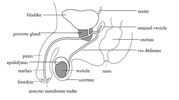
- Testes: The testes contain seminiferous tubules, where sperm cells are produced through cell division. The testes also store sperm and produce testosterone, a hormone responsible for the development of male secondary sexual characteristics.
- Vas Deferens: This sperm duct runs from the epididymis and serves as the pathway for sperm to travel from the epididymis to the urethra.
- Penis: Made of spongy erectile tissue, the penis is a sensitive organ that, when erect, delivers semen into the female's vagina, leading to fertilization.
- Prostate Gland: This gland secretes substances that help energize and transport sperm.
- Cowper's Gland: Secretion from this gland helps to normalize the alkaline concentration of sperm.
Female Reproductive System
The female reproductive system includes the following components:
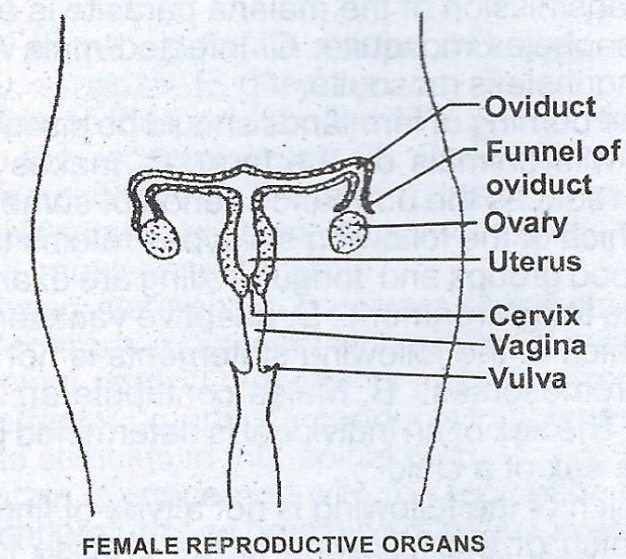
- Ovary: The ovaries contain thousands of potential eggs called primary oocytes. They are responsible for producing female gametes.
- Oviduct: The released egg enters a funnel-like structure known as the fallopian tube or oviduct, which transports the egg for fertilization. The oviduct connects to the uterus.
- Uterus: The uterus is a muscular structure connected to the exterior through the vagina. Its glandular lining nourishes the embryo during early development, and its smooth muscles increase in number during pregnancy. The uterus contracts during birth to expel the fetus and placenta.
- Cervix: Located at the lower part of the uterus, the cervix closes after fertilization to prevent further sperm and foreign bodies from entering.
- Vagina: The vagina receives sperm during intercourse and serves as the exit for blood during menstruation and babies during childbirth.
- Vulva: The external female genital organs. The vulva includes the inner and outer lips of the vagina, the clitoris, the opening of the vagina and its glands, the opening of the urethra, and the mons pubis (the rounded area in front of the pubic bones that becomes covered with hair at puberty).
The Egg
Female sex cells, or eggs (ova), are produced in the ovaries through a process called oogenesis. Human eggs are larger than sperm and consist of cytoplasm, a nucleus, granules, and yolk droplets. The yolk provides nourishment for the embryo, particularly during the early stages of development. The nuclei of both the sperm and egg contain chromosomes, which carry genes—the inherited traits passed from parents to offspring.
The Sperm
Male gametes, or sperm, are produced in the testes through spermatogenesis. A sperm cell is unicellular and consists of a head containing the nucleus, a middle piece, and a tail or flagellum.
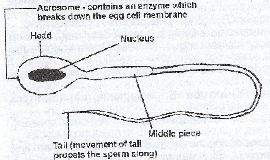
- Acrosome: Located at the head's posterior end, the acrosome contains enzymes that dissolve the egg membrane and facilitate egg penetration during fertilization.
- Nucleus: Found in the head, the nucleus carries the genetic material, which fuses with the egg’s nucleus during fertilization.
- Middle Piece: Contains mitochondria that generate energy for the sperm to swim toward the egg.
- Tail (Flagellum): The tail propels the sperm, enabling it to move toward the egg.
Menstruation
Menstruation is a cycle that involves changes in the uterus and ovaries, controlled by various hormones. This cycle typically lasts around 28 days:
At the beginning of the cycle, the lining of the uterus breaks down, resulting in menstruation. As follicles in the ovaries develop, the amount of estrogen produced by the ovaries increases. Estrogen acts on the uterus, causing its lining to thicken and develop more blood vessels. These changes help an early embryo implant if fertilization occurs. The pituitary gland in the brain releases follicle-stimulating hormone (FSH) and luteinizing hormone (LH), both of which help trigger ovulation.
Once the egg is released, the follicle that produced it turns into a solid structure called the corpus luteum. The corpus luteum produces a hormone called progesterone, which further thickens the uterine lining and encourages the formation of more blood vessels. If the egg is fertilized, the corpus luteum continues to produce progesterone, keeping the uterus in a state that supports embryo implantation. However, if the egg is not fertilized, the corpus luteum stops producing progesterone. This causes the uterine lining to break down, and the blood is shed, exiting through the cervix and vagina—this is the menstrual flow.